Procedures
Coronary Angiography
Coronary angiography is used to see how blood flows through the arteries in your heart.
Your doctor will give you instructions in order to prepare. The procedure is performed in an operating room called a cardiac catheterization laboratory. During coronary angiography, a local anesthetic is injected to numb the area where the catheter will be inserted. A very small incision is then cut in the skin over a blood vessel through which a small, thin tube (catheter) is threaded and the procedure performed. A small amount of dye is injected through the catheter to help with imaging during the procedure. You might feel pressure in the area where the catheter is inserted, but you shouldn't feel sharp pain. When the angiogram is over, the catheter is removed and the incision is closed. Following the procedure you will need to stay for observation and monitoring. You may be able to go home the same day, or you may have to remain in the hospital overnight.
Coronary Angioplasty and Stenting
Coronary angioplasty is used to open clogged heart arteries and coronary stenting supports the walls of your artery to help prevent it from re-narrowing after the angioplasty.
Your doctor will give you instructions in order to prepare. The procedure is performed in an operating room called a cardiac catheterization laboratory. During coronary stenting, a local anesthetic is injected to numb the area where the catheter will be inserted. A very small incision is then cut in the skin over a blood vessel through which a small, thin tube (catheter) is threaded and the procedure performed. A small amount of dye is injected through the catheter to help with imaging during the procedure. You might feel pressure in the area where the catheter is inserted, but you shouldn't feel sharp pain. A small balloon at the end of the catheter is inflated, widening the blocked artery in the effected area(s) and the spring-like stent expands and locks into place inside the artery. Because the balloon temporarily blocks blood flow to part of your heart, it's not uncommon to experience chest pain while it's inflated. Finally, the guide catheter is removed, and the procedure is completed. Following the procedure you will need to stay for observation and monitoring. You may be able to go home the same day, or you may have to remain in the hospital overnight.
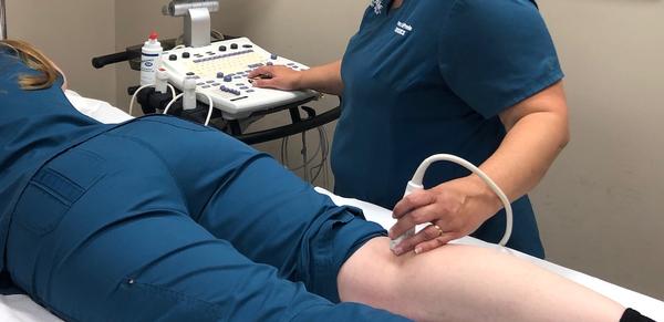
Peripheral Angiography
Peripheral angiography is used to find narrowed or blocked areas in one or more of the arteries that supply blood to your legs and lower body.
Your doctor will give you instructions in order to prepare. During peripheral angiography a local anesthetic is injected to numb the area where the catheter will be inserted. A very small incision is then cut in the skin over a blood vessel through which a small, thin tube (catheter) is threaded and the procedure performed. A small amount of dye is injected through the catheter to help with imaging during the procedure. You might feel pressure in the area where the catheter is inserted, but you shouldn't feel sharp pain. When the angiogram is over, the catheter is removed and the incision is closed. Following the procedure you will need to stay for observation and monitoring. You may be able to go home the same day, or you may have to remain in the hospital overnight.
Peripheral Angioplasty and Stenting
Peripheral angioplasty is used to open clogged peripheral arteries and peripheral stenting supports the walls of your artery to help prevent it from re-narrowing after the angioplasty.
Your doctor will give you instructions in order to prepare. During peripheral angioplasty and stenting, a local anesthetic is injected to numb the area where the catheter will be inserted. A very small incision is then cut in the skin over a blood vessel through which a small, thin tube (catheter) is threaded and the procedure performed. A small amount of dye is injected through the catheter to help with imaging during the procedure. You might feel pressure in the area where the catheter is inserted, but you shouldn't feel sharp pain. A small balloon at the end of the catheter is inflated, widening the blocked artery in the effected area(s) and the spring-like stent expands and locks into place inside the artery. Because the balloon temporarily blocks blood flow to part of your heart, it's not uncommon to experience chest pain while it's inflated. Finally, the guide catheter is removed, and the procedure is completed. Following the procedure you will need to stay for observation and monitoring. You may be able to go home the same day, or you may have to remain in the hospital overnight.
Inferior Vena Cava (IVC) Filter Placement
Inferior vena cava (IVC) filter placement is used to help reduce the risk of pulmonary embolism by trapping large clots and preventing them from reaching the heart and lungs.
Your doctor will give you instructions in order to prepare. During inferior vena cava (IVC) filter placement a local anesthetic is injected to numb the area where the catheter will be inserted. A very small incision is then cut in the skin over a blood vessel through which a small, thin tube (catheter) is threaded and the procedure performed. A small amount of dye is injected through the catheter to help with imaging during the procedure. You might feel pressure in the area where the catheter is inserted, but you shouldn't feel sharp pain. The IVC filter is then placed through the catheter and into the vein. Once it is in the correct position, the filter is released, allowing it to fully expand and attach itself to the walls of the blood vessel. Finally, the guide catheter is removed, and the procedure is completed. Following the procedure you will need to stay for observation and monitoring. You may be able to go home the same day, or you may have to remain in the hospital overnight.


